
Imagine being able to look inside your shoulder, knee, or back while you move. Not with a big MRI machine or a trip to the hospital, but right here during your PT session. That is exactly what musculoskeletal (MSK) diagnostic ultrasound allows me to do at JointWorks PT.
This technology has changed the way I evaluate and treat patients in Northbridge and Ashland. Instead of relying only on my hands and your description of pain, I can see what is happening beneath the skin in real time [1]. It takes the guesswork out of treatment and gives both of us confidence that we are targeting the right problem.
What Is MSK Diagnostic Ultrasound?
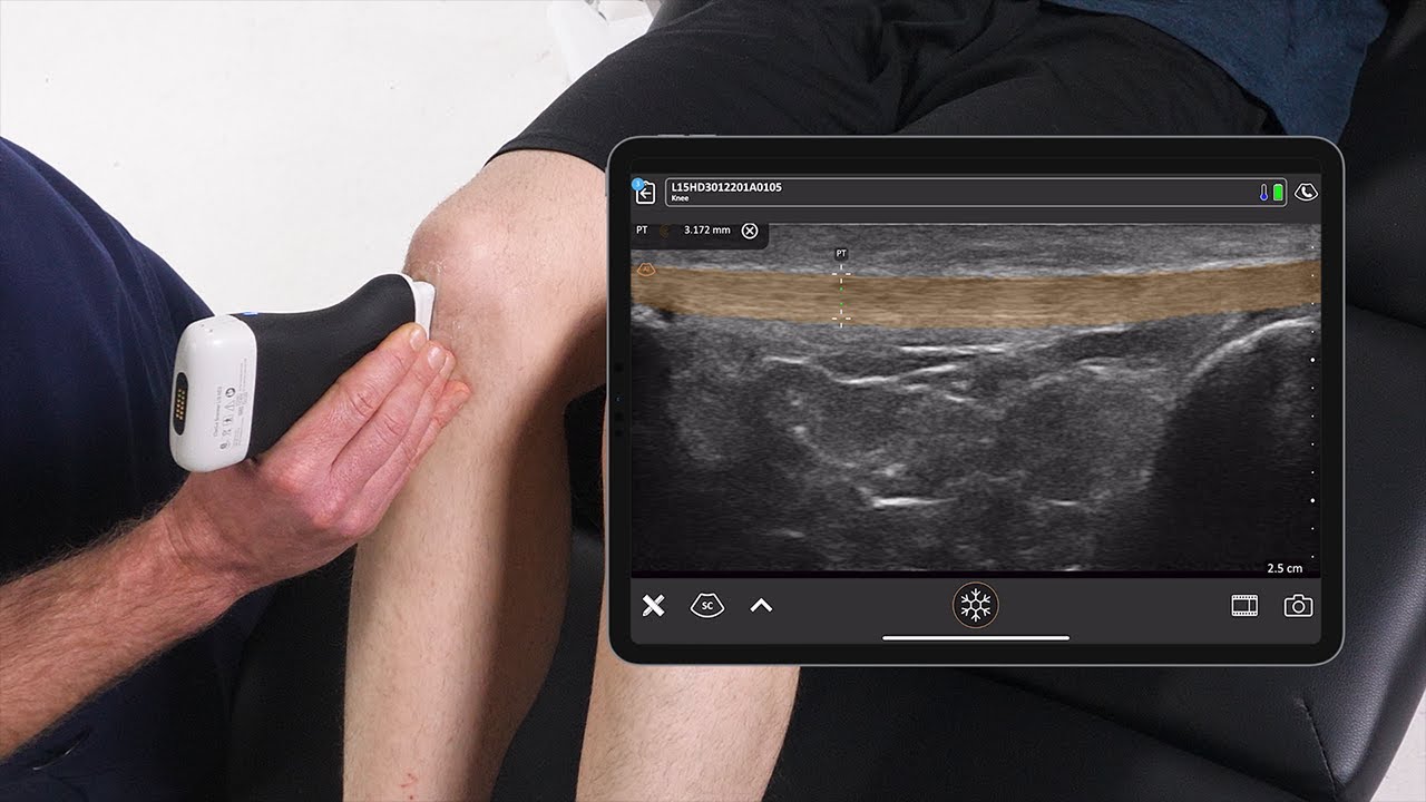
MSK ultrasound is the same type of imaging you might associate with pregnancy scans, but it is designed specifically for muscles, tendons, ligaments, and joints. A small handheld probe sends sound waves into your body, and the echoes create a live picture on a screen. There is no radiation, no injections, and no pain.
What makes this tool so valuable in physical therapy is the "real time" part. An MRI gives you a snapshot. Ultrasound gives you a moving picture. I can watch your tendon glide as you bend your elbow. I can see whether a muscle is firing properly as you do a specific exercise [2]. That kind of information is incredibly useful when building a treatment plan.
How I Use Ultrasound at JointWorks PT
I use diagnostic ultrasound in several ways throughout the treatment process.
Pinpointing the Problem
When you come in with shoulder pain, there are dozens of possible causes. Is it a rotator cuff issue? A swollen bursa? A problem with the biceps tendon? Ultrasound lets me look at each of these structures directly. Combined with my clinical exam, this helps me narrow down the source of your pain faster than guessing or waiting weeks for outside imaging.
Guiding Treatment Decisions
Once I know exactly what is going on, I can make better choices about your care. For example, if ultrasound shows that your Achilles tendon has thickening and disorganized fibers, that tells me we need a specific loading program to help those fibers remodel. Without that image, I might have treated it differently and seen slower results.
Tracking Your Progress
One of my favorite uses for ultrasound is showing patients their own healing. When you can see on a screen that your tendon looks healthier than it did four weeks ago, it builds motivation. Healing can feel slow when you are in the middle of it. Having visual proof that your body is responding to treatment makes a real difference.
Checking Muscle Activation
After surgery or a long period of pain, certain muscles can "shut off." They stop working the way they should. With ultrasound, I can watch a muscle contract (or fail to contract) while you do an exercise [3]. This is especially helpful for deep core muscles, the rotator cuff, and other areas that are hard to feel from the outside.
What Conditions Benefit from Ultrasound Imaging?
Diagnostic ultrasound is useful for a wide range of issues I see in the clinic every week:
- Tendon problems: Achilles tendon pain, rotator cuff issues, tennis elbow, and patellar tendon pain
- Muscle tears and strains: Seeing the size and location of a tear helps determine whether you need PT, rest, or a referral
- Swelling and fluid: Identifying joint swelling, bursitis, or fluid collections that may be causing pain
- Nerve issues: Checking for nerve compression or swelling in conditions like carpal tunnel syndrome
- Post-surgical recovery: Monitoring how tissues are healing after a procedure
- Chronic pain: When the source of lingering pain is unclear, ultrasound can reveal what other tests might miss
Why This Sets JointWorks PT Apart
Most physical therapy clinics do not offer diagnostic ultrasound. Typically, if your PT suspects a structural issue, they send you back to your doctor for imaging. That means more appointments, more waiting, and more time before you start the right treatment.
At JointWorks PT, I bring that imaging into the treatment room. You get answers faster. We adjust your plan sooner. And you can actually see what is happening in your body, which helps you understand your condition and stay engaged in your recovery.
I am currently pursuing my RMSK (Registered Musculoskeletal Sonographer) certification to further deepen my expertise in this area. This advanced credential will add another layer of formal training to the ultrasound skills I already use in practice. Combined with my board certification in orthopedics and fellowship training in manual therapy, diagnostic ultrasound is one more tool that helps me provide thorough, well-informed care.
What to Expect During an Ultrasound Assessment
If you have never had an MSK ultrasound before, here is what a typical session looks like:
- Quick setup: I apply a small amount of gel to the area being examined. The gel helps the probe make good contact with your skin.
- Live imaging: As I move the probe over the area, we both watch the screen. I will walk you through what we are looking at in plain language.
- Movement testing: I may ask you to move the joint or tighten a muscle so we can see how the structures behave during activity.
- No discomfort: The probe simply rests on your skin. There is no radiation, no needles, and no side effects.
- Immediate answers: Unlike an MRI, there is no waiting for results. We discuss findings right then and adjust your treatment plan on the spot.
The entire imaging process usually takes just a few minutes and fits naturally into your regular PT visit.
Ultrasound vs. MRI: When Each Makes Sense
Patients often ask me how ultrasound compares to MRI. Both are valuable, and they serve different purposes.
MRI is excellent for deep structures like the inside of a joint, spinal discs, and cartilage. It gives a detailed, static image that is useful for surgical planning.
Ultrasound excels at looking at tendons, muscles, ligaments, and nerves near the surface. Its biggest advantage is that it shows these structures while they move [4]. It is also faster, more accessible, and does not require a referral or a separate facility.
In many cases, ultrasound provides all the information I need to build an effective treatment plan. When deeper imaging is necessary, I will always refer you to the right specialist.
Technology That Serves the Patient
I did not add ultrasound to my practice because it looks impressive. I added it because it makes treatment better. When I can see exactly what is causing your pain, I can treat it more precisely. When you can see your own progress on a screen, you stay motivated. That combination of better information and better engagement leads to better outcomes.
If you are dealing with a stubborn injury, unexplained pain, or a condition that has not responded to other treatments, diagnostic ultrasound may give us the insight we need to move forward. I would love to show you what it can do.
References
- Bianchi S, Martinoli C. Point-of-Care Ultrasound, the New Musculoskeletal Physical Examination. PMID 33560035. PubMed
- Totten S et al. Exploring the Integration of Diagnostic Musculoskeletal Ultrasound Imaging into Clinical Practice by Physical Therapists. PMID 36259351. PubMed
- Teyhen DS. Rehabilitative Ultrasound Imaging Is a Valid Measure of Trunk Muscle Size and Activation During Most Isometric Sub-Maximal Contractions: A Systematic Review. PMID 19681737. PubMed
- Docking SI et al. Classification of Tendon Matrix Change Using Ultrasound Imaging: A Systematic Review and Meta-analysis. PMID 30007477. PubMed